


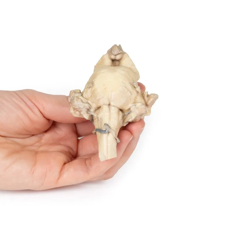

3D Printed Brain Stem, Isolated Anatomy From Midbrain to Medulla Oblongata
This 3D model provides a view of the isolated brainstem anatomy from the midbrain to the medulla oblongata, and
compliments the other diencephalon/brainstem 3D model (BR 10) in our series.
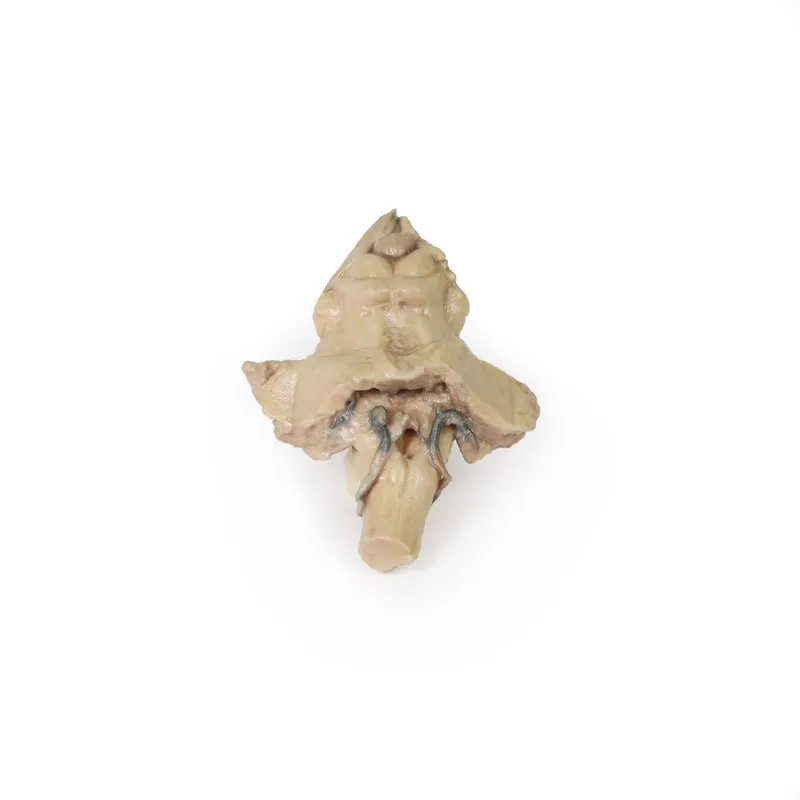
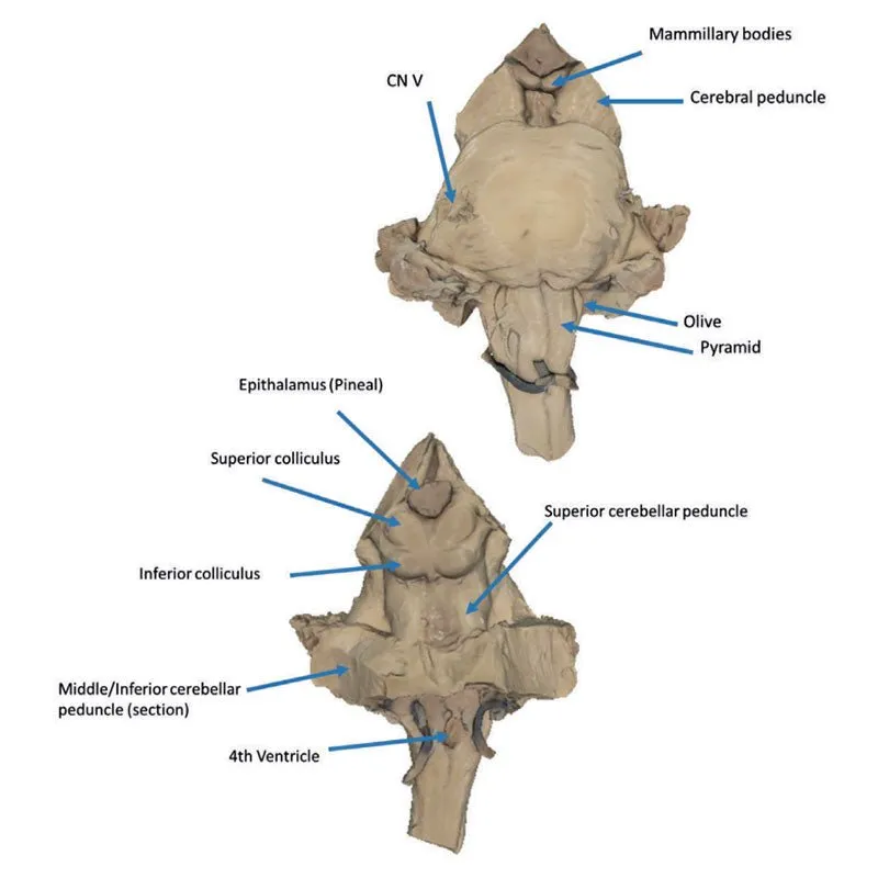
Rostrally, the 3D model has been
sectioned at an angle from the overlying diencephalon while retaining the mamillary bodies of the hypothalamus
between the cerebral peduncles (anteriorly) and the pineal gland/epithalamus (posteriorly). Posteriorly, the
corpora quadrigemina (the collective superior and inferior colliculi) of the midbrain are prominent adjacent to
the superior cerebellar peduncles. The cerebellum itself has been removed, leaving the cross-section of the
middle and inferior cerebellar peduncles on each side. Inferior to the sectioned peduncles is the partially
opened fourth ventricle and remnants of the posterior inferior cerebellar arteries.
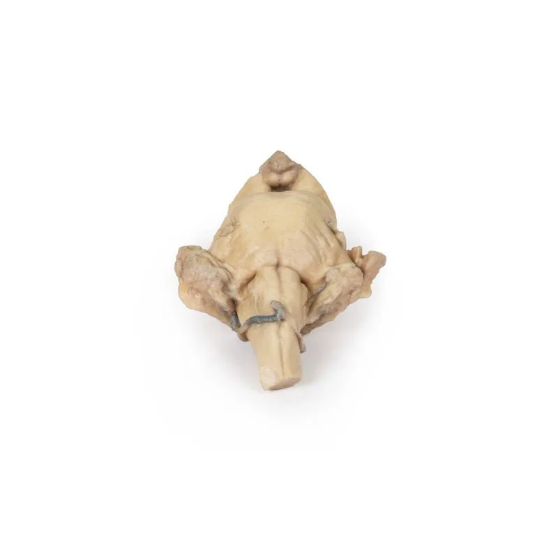
On the ventral aspect of
the 3D model the pons is preserved with the origin of the trigeminal nerve (CN V) preserved (particularly on the
left side). Inferior to the pons on the medulla oblongata, both the pyramids and olives are visible on both
sides (particularly clear on the right).
GTSimulators by Global Technologies
Erler Zimmer Authorized Dealer


